Charting sores and bruises in multiple colours
Detailed nuances in colour can reveal the age of bruises or detect when sores are not healing properly.
Denne artikkelen er over ti år gammel og kan inneholde utdatert informasjon.
Any parent can testify that kids cannot go through childhood without their share of bruises and scrapes. But not all of these injuries are so minor or innocent that a little consolation and a bandage will stop the tears.
Bruises – or contusions, injuries that don’t break the skin – can also be caused by domestic abuse or other crimes. It would help investigators if they could determine when a person was struck and how hard.
Nor do all cuts heal as well as an ordinary scraped knee. Many elderly people have poor blood circulation in their legs, and chronic lesions on their lower legs or feet need constant attention. Burns can also be hard to heal.
Multiple hues
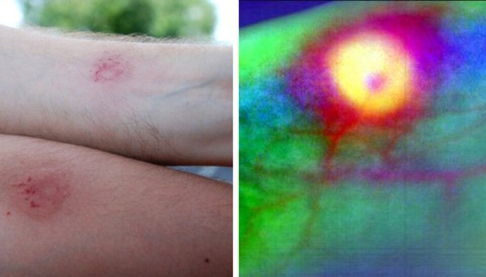
A hyperspectral camera can be helpful here. It can detect hundreds of different colour shades as compared to the common mixes of red, green, yellow and blue captured by a normal camera.
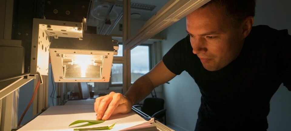
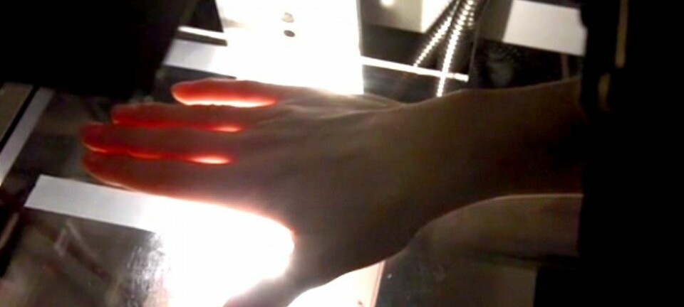
“We shine light on the skin and it penetrates. It is absorbed and spread, in part by blood and connective tissue. Some of this light is reflected back again and captured hyperspectrally. The nuances of colour in the reflected light can reveal a lot about sores and bruises,” says Lise Randeberg.
The hyperspectral camera is produced by the Norwegian firm Norsk Elektro Optikk. Lise Randeberg and her research colleagues have collaborated with scientists and doctors at St. Olavs Hospital in Trondheim in developing diagnostic methods that use the camera.
Paintballs and blunt objects
Randeberg is a professor of biomedical optics and photo techniques at the Norwegian University of Science and Technology (NTNU) and has used human volunteers who play with paintballs and anesthetized domestic pigs in her experiments with contusions.
Blows made by heavy blunt instruments were compared with light, quick hits. The heavy blows can be compared with punches from a heavyweight boxer. They resulted in haemorrhages deep in muscle tissue.
Paintballs strike lighter and faster when they hit. The damage they do can be compared to welts and bruises made by a flexible branch or a whip. Such injuries are more superficial.
Can see deep bruises
Powerful computing is needed to retrieve useful information from the camera. Hidden among hundreds of colour shades are the differences that interest Randeberg and her colleagues.
The data are first adjusted in keeping with the tone of the white light used in the photo. Then visual noise is filtered out and in some cases points in the picture are merged to find average values.
Specific points that have been selected as especially representative and colour variations that are independent of one another, and thus are most interesting, are accentuated.
Finally, the colour variations are compared with theoretical models of how light disperses through tissue in various kinds of injuries.
This is how Randeberg and her colleagues can create a picture of the injury that made blood accumulate, layer by layer, down into muscle tissue.
Shin sores and loose flaps of skin
“Another long-time project involves analyses of persistent sores,” says Randeberg. She has undertaken a number of tests, some with patients with chronic sores on their legs and others with small pieces of skin that were removed from different patients during plastic surgery and were subsequently donated.
The donated patches of skin were placed in a nutrient solution that keeps skin cells alive. Then the researchers created a sore in the centre of the patch of skin tissue.
Hyperspectral images and tissue samples were taken of the skin patches every third day for three weeks. During most of this period the sore healed on the living patches of skin.
Data from the first period was used as a basis for comparison in order to monitor the changes when the sore grew on the patches of skin. The lesion on the patient with a lower leg sore was also evaluated at intervals by a skin doctor to compare the results.
Could see where the growth occurred
After the data was cleaned up the scientists could see changes in the nuances of colour in the hyperspectral images.
This enabled them to discern between uninjured skin around the sore, newly produced skin in the region that had healed and areas of the sore that had not healed.
In one of the tests the scientist took 3D pictures with two normal cameras to chart the depth of the sore. This enabled them to quantify the amount of tissue that was still missing in the lower leg sore.
“Doctors are interested in knowing how the volume of the sore changes in the course of time,” says Randeberg.
Important tool
“Hyperspectral photography can be a quick way of monitoring wounds without needing to take samples of the sore tissue,” she says. “Nearly a third of all amputations are due to persistent sores that won’t heal. So this can become an important medical tool.”
----------
Read the Norwegian version of this article at forskning.no
Translated by: Glenn Ostling
External links
- Characterization of vascular structures and skin bruises using hyperspectral imaging, image analysis and diffusion theory, Journal of Biophotonics No. 1-2, pp. 53-65, 2010, DOI 10.1002/jbio.200910059
- Hyperspectral Imaging of Bruises in the SWIR Spectral Region, Photonic Therapeutics and Diagnostics VIII, Proc. of SPIE Vol. 8207, 2012, sammendrag, doi: 10.1117/12.909137
- Hyperspectral imaging as a diagnostic tool for chronic skin ulcers, Photonic Therapeutics and Diagnostics IX, Proc. of SPIE Vol. 8565, 2013, doi: 10.1117/12.2001087
- Combined Hyperspectral and 3D characterization of non-healing skin ulcers, Colour and Visual Computing Symposium, IEEE Xplore 2013, doi 10.1109/CVCS.2013.6626271
- Hyperspectral characterization of an in vitro wound model, Photonic Therapeutics and Diagnostics X, Proc. of SPIE Vol. 8926, 2014, sammendrag, doi: 10.1117/12.2039976
- Skin changes following minor trauma, Lasers in Surgery and Medicine, sammendrag, Vol. 39 Issue 5, s. 403-413, 2007, DOI: 10.1002/lsm.20494


