Diagnosing arthritis with a colour scan
An easily used screening tool shines new light on how to detect arthritic inflammations.
Denne artikkelen er over ti år gammel og kan inneholde utdatert informasjon.
Arthritis is a swelling of the joints that causes stiffness and pain. As time passes the inflammation of stricken joints can be crippling.
Arthritis often starts in the hands and feet. These appendages can be diagnosed by a new tool which detects arthritis by shining light through the skin, blood vessels, muscle, tendons and joints.
Fine nuances of colour
“Fingers and toes are thin enough to shine light through,” says Lise Randeberg. She is a professor in biomedical optics and photonics at the Norwegian University of Science and Technology NTNU.
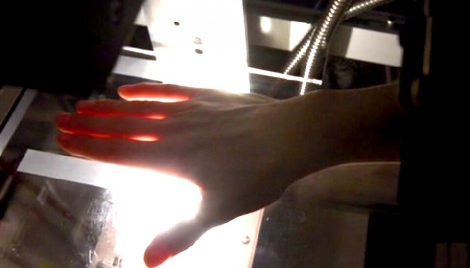
You may have tried it with a strong flashlight. When white light passes through a hand we can see red light emerging from the other side. Randeberg and her colleagues have put this to good use in the method they are developing. Small nuances of colour and other changes in the light can reveal the presence of arthritis.

This gives an easy and immediate diagnosis. More people can be examined and new cases can be detected early, before the disease cripples joints for good.
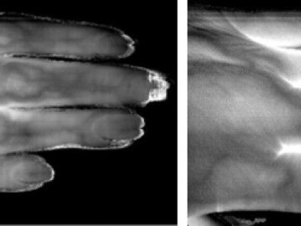
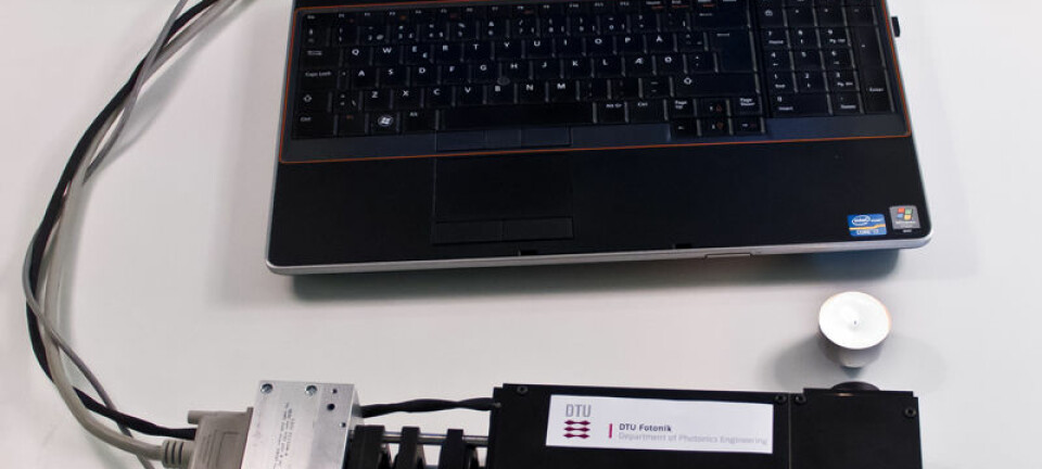
A subject’s hands are scanned by a hyperspectral camera developed by the firm Norsk Elektro Optikk. It builds up an image line by line, like a table scanner, and can discern among many more nuances of colour than do normal scanners or cameras.
Seeing different kinds of arthritis
“We have done preliminary tests in cooperation with rheumatologists at St. Olav Hospital in Trondheim,” says Randeberg.
“We transilluminated the hands of 15 patients with arthritis and 20 healthy persons. We found that the scannings might also be able to differentiate between different types of arthritis. They results we got were unexpectedly good,” she says.

Although the images show clear differences, these have to be interpreted to give accurate diagnoses. Randeberg and her colleagues are now developing computer models which can do just that. But it’s a challenging task.
Into the depths
“When light passes through a hand we are especially interested in indications from the blood. Inflammations create changes in blood circulation and this is something we can quantify,” explains Randeberg.
But measurements of blood circulation alone do not indicate different types of inflammations. These can have other causes than arthritis. They can be due to a physical injury or an infection. To determine the signs of arthritis, medical personnel need to get a picture of exactly where the blood is and how much oxygen it contains.
The hyperspectral camera takes a picture that provides lots of detailed nuances of colour. But this isn’t sufficient on its own for arriving at accurate diagnoses. The medical scientists need to see what is happening from top to bottom through the hand or foot. They need to see in depth, in three dimensions. How do they do it?
Plenty of computing power
“We make models of how the light moves through blood, muscles, ligaments and joints. When such models are linked to the images from the hyperspectral transilluminations, the scientists can interpret information from the depths as well. But this requires a lot of computing power.”
“We are making every effort to get a completed computer analysis within a minute after the transillumination. It doesn’t help if the transillumination is quick and easy if the patient still has to wait weeks for a result. If such a delay were okay, other methods would work about as well,” says Randeberg.
Dilemma in blue and green
Another challenge is that light through the hand is subdued greatly by blood and skin pigments. This makes the nuances of colour harder to catch with the hyperspectral camera.
Blue and green light is reduced the most, especially among people with dark skin. This is also the colour range where the interesting signals from blood is the strongest and light is the weakest. This is a dilemma. For the method to work universally it needs to function with all our human skin colours.
This means that researchers need to compromise on the colours they utilise in the analysis. The further into the red range of the spectrum, and further into higher energies and what for our eyes is invisible light – infrared – the more light makes it through the hand. This in turn weakens the nuances of colour that can be detected from blood.
EU cooperation
The solution is a new hyperspectral camera in which the colour spectrum is shifted somewhat from visible light toward infrared. This camera has been developed specially for medical research by Norsk Elektro Optikk.
“We are now going to test this camera at a clinic in Germany. The project involves collaboration with, among others, the German Fraunhofer Institute, Norsk Elektro Optikk and NTNU, and is financed by the EU,” says Randeberg.
Nearly ten years have passed since the first time Randeberg placed her hand beneath a hyperspectral camera from Norsk Elektro Optikk.
“Until then we had worked at seeing the colour from light at one spot. It was really a new world for us to chart the nuances of colour in a hyperspectral analysis. We were also among the first to use hyperspectral analyses in medical research.”
“Much has changed since then. We started in the blue, and now we see in red – and infrared. This is really brilliant,” says Randeberg.
----------
Read the Norwegian version of this article at forskning.no
Translated by: Glenn Ostling


