Do you have a greater risk of breast cancer if your breasts are big or small?
ASK A RESEARCHER: Risk is tied to the amount of breast tissue, cancer researcher says.
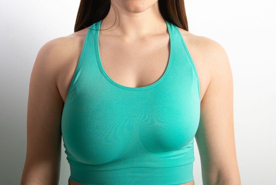
One of our readers says that she has quite small breasts. She wonders if it makes a difference in her risk of getting breast cancer.
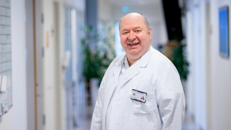
We asked Håvard Søiland, a professor of surgery at the University of Bergen and a senior researcher at Stavanger University Hospital.
“This is a question that has been discussed time and again in professional circles,” Søiland says. He has studied breast cancer for 20 years.
No one knows the right answer.
Nevertheless, Søiland can say a lot about which women and which breasts have a higher risk of developing breast cancer.
Tumours most often in the upper, outer quarter
Tumours occur more often in a specific part of the breast.
Imagine your chest is a clock. The nipple is in the middle of the clock. 12 is the top of the chest and 6 is straight down. 3 is towards the armpit. 9 is towards the middle of the chest.
Women more often get tumours in the area between 12 and 3.
This pattern ultimately led researchers to a dead end.
“For a period, people began to suspect that deodorants contained substances that penetrated the skin. This was refuted by several large studies. But this dead end actually led researchers to learn more about breast cancer,” Søiland tells sciencenorway.no.
This quarter circle of the breast has more tissue. And the more breast tissue, the greater the risk of cancer.

Easier to find tumours
Regardless of breast size, women have a greater chance of getting cancerous tumours in this area.
Those with small breasts still have an advantage.
“Women with small breasts find it easier to detect tumours. That means they have a better prognosis," Søiland says.
Large breasts make it more difficult to detect tumours before they become large.
No definite connection
There has been a lot of research into whether the size of a woman’s breasts results in a greater chance of breast cancer. Søiland highlights one of several summaries in which researchers looked at studies from 1950 to 2010.
“They did not find any reliable results that show a connection between breast cancer and breast size," he says.
Nevertheless, some women with large breasts do get breast cancer more often.
Tall women have a 17 per cent higher risk.
The increased risk is due to tall women having a greater amount of the growth hormone HGH, which stands for human growth hormone.
Some women choose to make their breasts smaller through plastic surgery. Several studies show that this affects cancer risk.
“Surgery removes breast tissue. So these women have a 20 per cent lower risk of breast cancer,” Søiland syas.
Density is more important
If you have large breasts, you don’t automatically have more breast tissue.
“Large breasts often contain more fat, not more breast tissue,” Søiland says.
One factor that is more important than size is the density inside the breasts. Radiologists have determined this through mammography.
“What we call dense breasts, meaning those with a lot of connective tissue and breast tissue, have a greater risk of developing cancer,” Søiland says.
That leads us to the question: What causes some women to have more breast tissue than others?
Why more or less breast tissue?
There are simply genetic differences in how much breast tissue women have. In addition, the female hormone oestrogen plays a role.
Søiland explains:
The mammary gland develops in the foetus in week six. This gland is influenced by the mother's oestrogen. The amount of breast tissue that is formed is related to the amount of hormonal stimulation from the mother.
Babies are also hormonally stimulated with oestrogen through breastfeeding.
Then comes puberty. Women develop different amounts of oestrogen during this time.
“Oestrogen is the big, ugly wolf when it comes to breast cancer,” says Søiland.
Reset during pregnancy
Oestrogen both initiates and allows breast cancer to develop.
The risk of breast cancer is related to how many years your body is affected by oestrogen.
“When women are pregnant, their oestrogen level is reset to zero. You get a break,” Søiland says.
Breastfeeding reduces the risk of breast cancer by 20 per cent.
In the old days, when women had 10 children, they had almost no breast cancer. Whereas nuns in convents have a much higher risk.
A woman with small breasts, who has not been pregnant or who gives birth late, probably has a larger risk of breast cancer than a woman with larger breasts who has given birth to children, according to the cancer researcher.
Women who have an abortion also have their oestrogen levels reset.
Number of years with oestrogen
Many women choose to take oestrogen supplements during menopause. This increases the risk of breast cancer.
“The reason is that it increases the number of years the breasts are exposed to oestrogen and synthetic progesterone,” says Søiland.
200 out of 3,600 cases of breast cancer can be explained by the use of hormone supplements after menopause, according to one study.
After menopause, the amount of breast tissue decreases. The breasts then consist mostly of fat and connective tissue.
Paradoxically, the risk of breast cancer increases from 50 to 70 years of age.
“The reason for that is that the breast tissue has been influenced by accumulated oestrogen for many years. The effect of this influence makes itself felt,” Søiland says.

Just bad luck
Søiland emphasises that this is only true for ordinary breast cancer. Consequently, this does not apply to the two per cent of breast cancer cases that are caused by an inherited gene called BRCA.
“Ordinary breast cancer is all about bad luck,” says Søiland.
You can't feel your breasts to tell if you have a lot of tissue or a lot of fat. But radiologists can see that on mammograms.
Solveig Hofvind heads BreastScreen Norway, which is part of the Cancer Registry of Norway.
She prefers to talk about glandular tissue.
“Breast tissue includes glandular tissue, connective tissue, supporting tissue and fatty tissue,” Hofvind tells sciencenorway.no. “Here we are very concerned with dense glandular tissue. A lot of research has been done on this, but there is still a lot we don't know."
White and light grey glandular tissue
On mammography images, the glandular tissue looks white and light grey. Fat tissue is black or dark grey.
But it's still not that simple. Because the gland density in the breast affects the images.
Researchers at the Cancer Registry of Norway have measured gland density in 400,000 mammography examinations using artificial intelligence. They then analysed the risk of developing breast cancer.
“Women with dense glandular tissue have an increased risk of breast cancer,” says Hofvind.
In addition, it is more difficult to find tumours in breasts with dense glandular tissue.
The parrot in the tree
Hofvind compares a breast with a cancerous tumour to a birch tree containing a parrot.
“In the middle of summer, the tree is full of sap and has lots of lush, green leaves. It is not so easy to see the parrot sitting in the tree,” she says.
In autumn, the sap flows out and the leaves disappear. In the breast, glandular tissue is replaced by fat.
“Then it’s easy to see the parrot. In the same way, it’s easier to see a cancerous tumour in a breast with the most fat,” says Hofvind.
Young women have denser glandular tissue than older women. Breast tissue density decreases, especially after menopause. Women who take hormones during menopause therefore maintain a certain glandular density.
Radiologists have measured breast tissue since the dawn of time, according to Hofvind.

You won't get definite answers
“These are subjective measurements. Therefore, you may be told you have one density when screened in one place and another density in another place. Many studies have shown this,” Hofvind says.
It’s possible to introduce a system for density measurements for all health institutions in Norway.
“We could have achieved that. Artificial intelligence will be able to provide objective values for mammographic density,” Hofvind says.
Even if you learn that you have dense glandular tissue, the problem is not solved. Because what are you going to do next?
Studies from BreastScreen Norway show that around five per cent of women have extremely dense glandular tissue. However, they do not undergo additional examinations.
“There are not enough resources to offer everyone with dense glandular tissue additional examinations using an MRI. We would also run the risk of doing a lot of unnecessary investigations by getting a lot of false positive findings,” Hofvind says.
There are now new European guidelines recommending extra measures for women with dense glandular tissue.
“There are several exciting studies underway on offers for these women, including the use of artificial intelligence in screening,” she says.
Size does not count in treatment
When it comes to breast cancer treatment, size doesn't matter.
“There is little difference in the prognosis if a woman has large or small breasts,” Søiland says.
It can be easier to do surgery on larger breasts, but when it comes to radiation, there is no difference. The specialists are very accurate. According to Søiland, Norway is advanced when it comes to radiation research.
What matters for whether you survive breast cancer is which biological subtype of cancer tumour you have developed. Some tumours are simply more dangerous than others.
“That is why we now carry out examinations of each tumour, in order to determine the inherent characteristics of the cancerous tumour. The treatment is then adapted to the tumour,” Søiland says.
This is what is called personalised treatment, which is now common for cancer patients.
Søiland himself conducts research on cancer recurrence.
“I have studied whether there are signs in the blood that warn of relapse before we can see it in other samples,” he says.
His research has succeeded in showing signs of recurrence a year before the tumour is detected by other means. But there is still a long way to go before this comes into regular use in hospitals.
“It can take up to ten years, but we are researching this further. We could then start treatment in round two of cancer earlier than we do today,” Søiland says.
———
Translated by Nancy Bazilchuk
Read the Norwegian version of this article on forskning.no


