
Annonse
Tumours in breasts with dense tissue are more difficult to detect. Artificial intelligence can simplify the job, a new study shows.
The new study is based on screening using an MRI. But women with dense breast tissue who are covered by the Norwegian mammography programme are not offered this type of screening.
Published
A woman's breasts are usually mostly fat, along with mammary glands.
But some women have breasts with much more glandular tissue than others. The glandular tissue is generally called dense breast tissue. Women with this kind of breast tissue have a higher risk of breast cancer.
It is also more difficult to detect cancerous growths in this kind of tissue using conventional mammography.
Fewer helping hands in the future
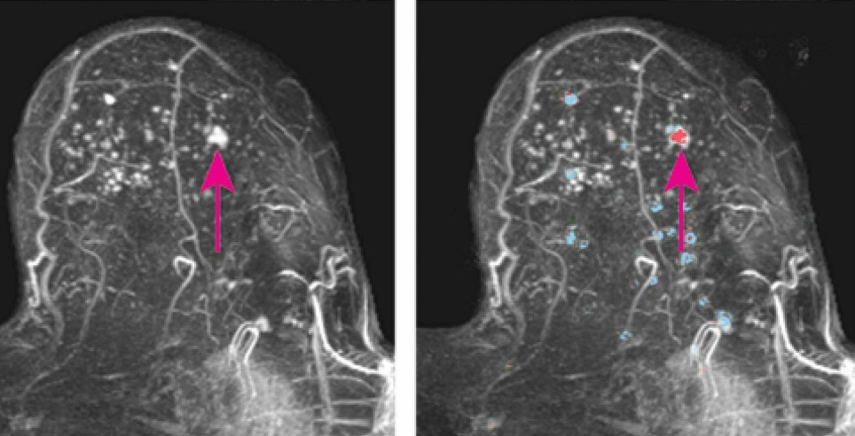
MRI scans provide more detailed images than conventional mammography. At the same time, it takes a lot of time for radiologists to go through MRI images to assess them for tumours.
Annonse
Now, researchers at a university hospital in the Netherlands have shown that artificial intelligence (AI) can do some of this work.
“We know that there will be fewer hands available per patient in the health care system in the future, which makes it important to find methods that enable us to solve our tasks with fewer resources,” Solveig Roth Hoff wrote in an email to sciencenorway.no.
Hoff is a section chief physician at Ålesund Hospital and studies imaging techniques that can detect breast cancer at the hospital and the Norwegian University of Science and Technology, NTNU.
Pick out breasts without tumours
More than 4,500 women participated in the Dutch MRI study. All had extremely dense breast tissue.
The task for the artificial intelligence was to pick out breasts without tumours.
The idea is that AI will save doctors from having to look through thousands of images of healthy breasts. That leaves them more time to concentrate on women who may have cancer.
A good start
Artificial intelligence, which is basically a computer program, identified 40 per cent of the healthy breasts.
And none of the breasts that actually contained cancerous tumours were misclassified by the program.
“The results were better than expected,” said Erik Verburg at University Medical Center Utrecht in a press release. Verburg is one of the researchers behind the new study.
“Forty per cent is a good start. At the same time, we still have sixty per cent to improve,” Verburg said.
Practiced on images from seven hospitals
The intelligent computer program was set up so that it could practice with some MRI images. It had to try to use what it had learned to assess the rest.
Annonse
The study included MRI images from eight hospitals.
The artificial intelligence practiced recognizing normal breasts in images from seven of the hospitals.
The computer program relied on the assessment of the images that doctors had made. After the artificial intelligence had trained itself, it tested itself on images from the eighth hospital.
Then the whole procedure was repeated seven more times, so that each hospital was used to test the program.
No MRI for dense breast tissue in Norway
But even though this approach is a common way to create computer programs that can teach themselves, it’s not a given that it will work as well with other MRI images.
“It is important that artificial intelligence programs be properly tested and validated before they are available for commercial use. This kind of program has to be tested on several different types of data sets from different hospitals, machines and populations,” Hoff said.
In Norway, however, it is not common for women with dense breast tissue to be referred for an MRI examination.
Applies to only five per cent
Nataliia Moshina has investigated whether Norway’s mammography programme should be changed so that women with dense breast tissue receive more follow-up.
One change could involve offering these women more frequent mammography screening or offered other kinds of screening, such as MRI.
Moshina concluded in her doctoral dissertation in 2017 that it’s too early for Norway to make this change.
First, doctors need a better method to figure out which women actually have dense breast tissue, she said.
Her research indicates that only five per cent of women who are screened in Norway have breasts with dense tissue.
Could have been useful for another group
Some women in Norway are already being offered MRI screening for breast cancer. These women generally have congenital genetic problems that give them a much higher than average risk of breast cancer.
“This is not such a very large population, but a (AI) program that could enable us to use fewer resources on this patient group would be useful, of course,” Hoff said.
At the same time, it is not certain that the Dutch computer program would have worked on breasts from this group.
To know if this screening approach would work for women with a predisposition to breast cancer, researchers would have had to test the program on these women in their study, Hoff said.
Reference
Erik Verburg et al.: Deep Learning for Automated Triaging of 4581 Breast MRI Examinations from the DENSE Trial, Radiology 10/2021


