
Annonse
Share your science:
The beauty of a swirl: We continuously reveal the secrets of the heart
SHARE YOUR SCIENCE: The heart is perhaps the most romanticised, and most studied human organ. The blood flow in the heart has fascinated physicians and researchers for decades, and our understanding of the cardiac mechanics are ever evolving.

Published
For the past 70 years, physicians have
used ultrasound in the examination of the heart, and these seven decades have
given us an enormous development of the method. Ultrasound of the heart, or echocardiography,
is a complex examination as it involves moving tissue and muscle and moving
blood.
Already in the 16th century Leonardo da Vinci described the possibility of swirls of blood, or vortices, in the aorta above the aortic valve.
Since the 1970’s we have used the Doppler technology to get an impression of the blood flow, and even though the ultrasound probe and scanner has evolved, the theory behind is the same today as was used 50 years ago.
Soundwaves and the Doppler effect
Sound is pressure variations and propagates as waves – and this is why we talk about sound waves or pressure waves. The frequency is the number of oscillations per second of the sound wave.
Imagine the sound of an ambulance as it moves towards you and the change in the siren as it passes and moves away. This is the Doppler effect. More specific the Doppler effect is the change in frequency of a soundwave when the source of the wave, i.e. a siren, is moving relative to the person that perceives the sound.
Annonse
In echocardiography, we utilise the Doppler effect to estimate velocities of both the contracting and relaxing cardiac walls and the moving blood pool within, as these move towards and away from the ultrasound probe.
The ultrasound wave is reflected towards the probe when it hits blood and tissue, and when these are in motion the Doppler effect arises with different frequencies of the waves that are transmitted and received, and this difference is measurable!
The Doppler technology is heavily limited by the angle of the blood relative to the ultrasound beam.
To be sure that our measurements are accurate, we are dependent on the ultrasound beam being well aligned with the flow direction, if not we will get a measurement of the velocity of the blood that is lower than the real velocity. And if the angle is too big, we will not get a measurement at all.
Flow perpendicular to the beam is impossible to interpret and flow that is not aligned with the beam is a challenge as well.
Visualizing complex blood flow is therefore not possible with the Doppler technology, and clinicians are dependent on interpretation rather than objective measurements.
Vortices and da Vinci
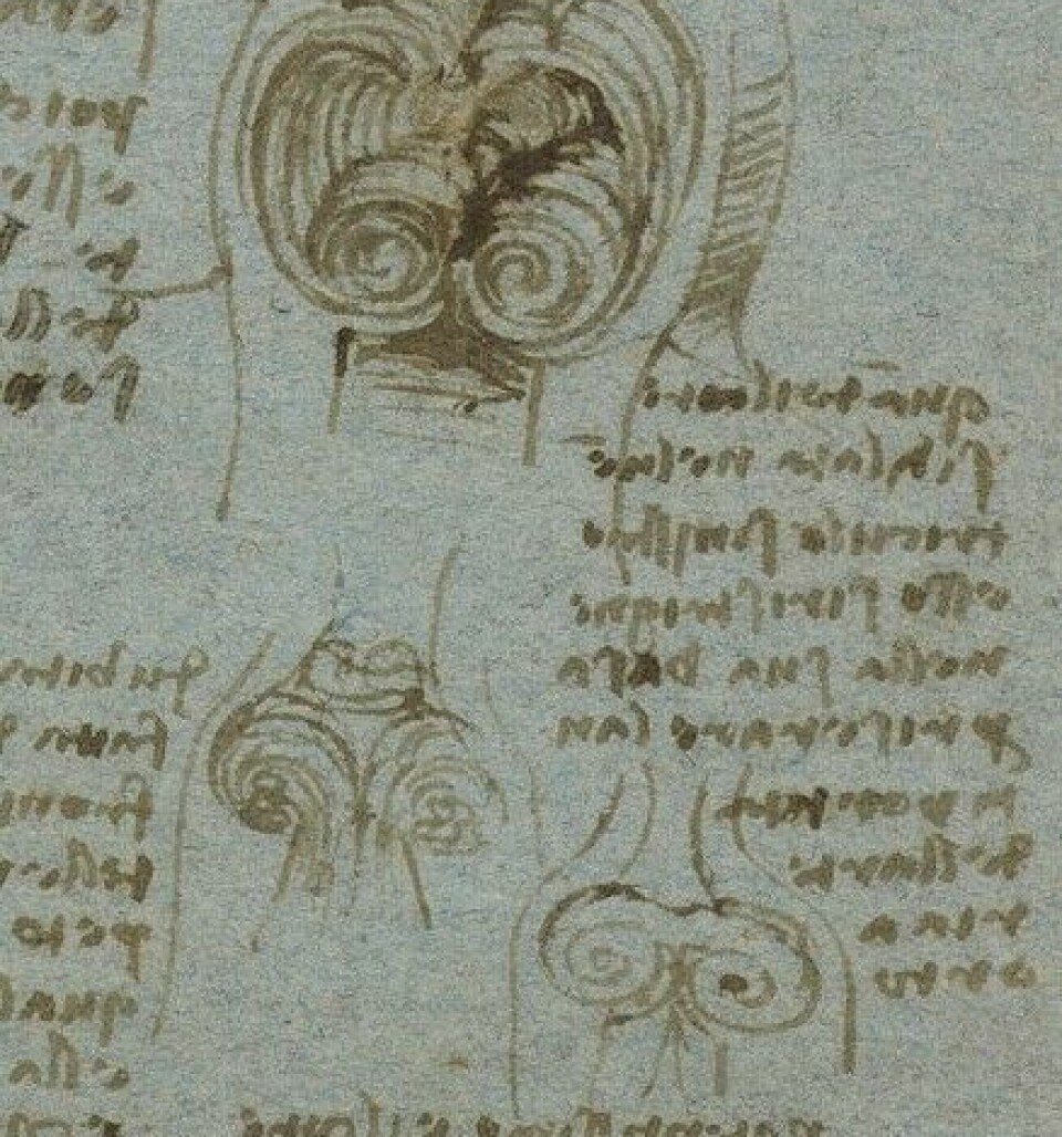
Already in the 16th century Leonardo da Vinci described the possibility of swirls of blood, or vortices, in the aorta above the aortic valve.
During the last 50 years researchers have dug deeper into this, and with fluid mechanics and models of the heart it has been shown that the anatomy of the heart will create a more complex blood flow than what we can see with Doppler technology alone.
The role of the vortex in normal heart function is to stop the blood colliding with itself, thus preserving the momentum and energy, and avoiding unnecessary loss of pressure and speed of the blood flow.
There is extensive research throughout the world today on different methods for vortex imaging and parameters linked to the formation of vortices in the ventricles of the heart.
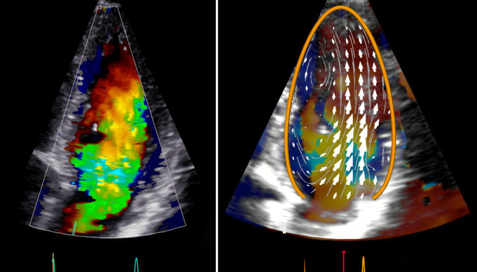
Tracking the blood
In 2018, we conducted a study in healthy volunteers to test a new method, first developed for kids, in adults. The method is called Blood Speckle Tracking (BST), and with this we can measure the movement of the blood in all directions inside the heart.
As opposed to the Doppler technology it is not dependent on the angle of the ultrasound beam. The motivation was to be able to visualise complex blood flow and vortices with ultrasound.
Annonse
Blood Speckle Tracking is promising as an addition to the standard clinical methods in children, and the aim of the study was to further develop the method so that it could be used in adults.
The challenge in adults when compared to children is that the heart is located deeper into the body, and that the ultrasound waves thus must penetrate deeper into the tissue.
We managed to adapt the method for adults, and from the study we could map the blood flow throughout the whole heart contraction (known as the cardiac cycle), describing the function of the blood flow and how it relates to the appearance and movement of the inside of the heart.
The results show a fascinating swirling pattern inside the heart, helping the muscle of the heart during the cardiac cycle so that the heart avoids using excessive force to pump blood to all parts of out body, thus conserving energy.

Changes in the blood flow
To better understand the normal physiology of the blood flow within the heart will in turn give a better understanding of how this will change with different heart diseases, and in 2020 we conducted a study in patients with heart disease.
In this study we had two groups of patients with heart failure. One of the groups had a dilated left main chamber with weakened muscle and impaired function, and the other one had thickened and stiff muscle which leaves little room for the blood in the left main chamber.
The first results from this study give us the impression is that the blood flow is changed, and that the vortices change with different heart diseases.
Our hope for the BST-method is that it can be a supplement to the Doppler technology, and that the methods together can help us better understand the spectrum of heart diseases. Maybe the method can be used to target medical treatment of heart failure – to fix the broken swirl.
FURTHER READING:
Share your science or have an opinion in the Researchers' zone
The ScienceNorway Researchers' zone consists of opinions, blogs and popular science pieces written by researchers and scientists from or based in Norway.
Want to contribute? Send us an email!


