Annonse
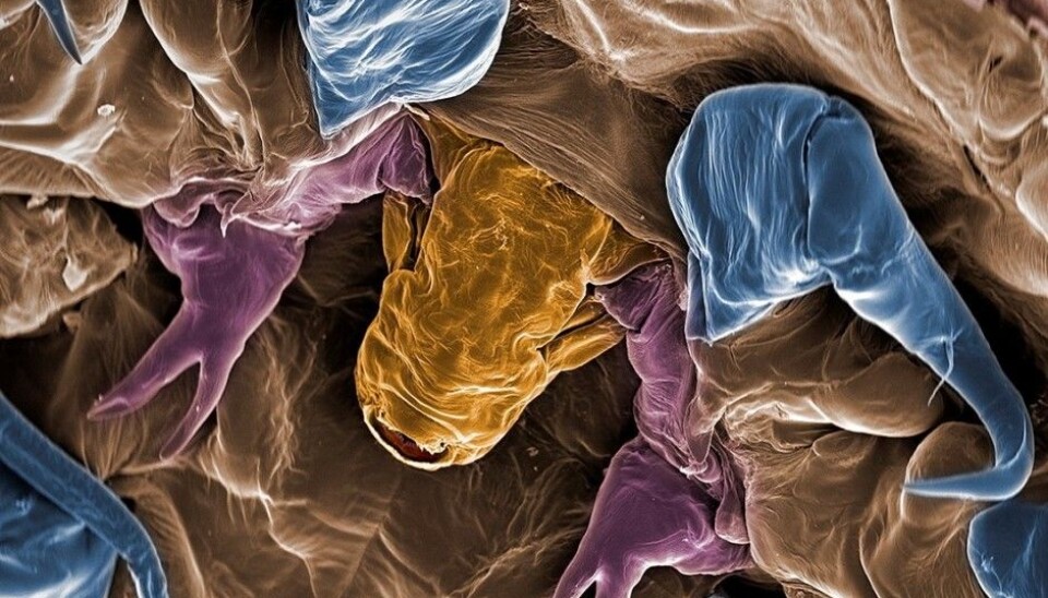
This is the face of a salmon louse
Most things on our planet are too small for us to see. Powerful microscopes give scientists the opportunity to catch a glimpse of what we are missing. Check out these close-ups of some of the micro-life in our midst
This summer the Science Park at the Norwegian University of Life Sciences (NMBU) in Ås exhibited some unique photos in an exhibition titled “See into the invisible”. These have been taken down now, but here are some of them.
Salmon louse
The scientific (Latin) name of this creature is Lepeophteirus salmonis. This parasite lives in salt water on salmon in the Northern Hemisphere. The louse has a diameter of 3–5 millimetres. These pictures have been coloured.

(Photo: Jannicke Wiik-Nielsen at the Norwegian Veterinary Institute.)
Annonse
A closer view below clearly reveals the mouth of the salmon louse.

(Photo: Jannicke Wiik-Nielsen at the Norwegian Veterinary Institute.)
Bee guts
This picture is of the intestinal tract of a honeybee. The blue area is the mid-stomach; the red is the ileum, the last part of the small intestine. They yellow part is a bee rectum. The photo was taken with an electron microscope at the NMBU Imaging Centre, coloured and magnified at a power of 70x.

(Photo: Åsmund Andersen in connection with a master degree thesis. It was reproduced and coloured by Odd Andersen.)
Pores of a leaf
The photo below is of stomata, the pores of leaves. These openings are vital to photosynthesis. Air, with its carbon dioxide and oxygen, enters and exits the plants through the stomata. Plants take carbon dioxide in and as a by-product of photosynthesis they release oxygen through these openings.
There can be as many as 30,000 of these openings per square centimetre on the undersides of leaves. Most plants have more stomata on the undersides of their leaves than on the top sides.

(Photo: Taken by Hilde Kolstad, senior engineer at the Imaging Centre of NMBU and coloured by Egil Paulsen).
Amoeba
Perhaps you have heard amoeba used as a derogatory description of someone? Do you really know what one is and how it looks?
Annonse
Amoebas are single-celled organisms about a half millimetre in size. They live in the sea and in fresh water, moist soil and as parasites in animals, including an estimated 50 million people, killing some 100,000 a year.
Beneath is a Paramoeba perurans. It causes a gill disease in fish and is a problem for the aquaculture industry.
The photo was taken with a scanning electron microscope and magnified several thousand times before being colour treated.

(Photo: Jannicke Wiik-Nielsen at the Norwegian Veterinary Institute. She used an electron microscope at the NMBU Imaging Centre)
Ringed Tubularia
It resembles a flower but this carnivorous critter belongs to the animal kingdom and is classified as a hydrozoan, which includes jellyfish and corals.
It has a mouth and uses the tentacles to catch plankton and other small victims that float by. This grows in seawater on solid surfaces such as buoys, mussels, ropes, rock and the hulls of boats.
The ringed tubularia below is about one centimetre in size.

(Photo: Jannicke Wiik-Nielsen at the Norwegian Veterinary Institute)
Ringed tubularia, or Ectopleura larynx, can clog the pens of aquaculture facilities – fish farms – and researchers have experimented with getting rid of them with ascetic acid, think vinegar. The photo below shows a ringed tubularia that has been subjected to this sort “acid test”. It has protected its reproductive organs by embracing them with its tentacles.
The photo was taken with a scanning electron microscope and magnified several hundred times. It has also been colour treated.

(Photo: Jannicke Wiik-Nielsen at the Norwegian Veterinary Institute)
Cross-section of a wheat root
The photo below was taken through a microscope and shows a cross-section of the root of wheat, the spongy tissue called aerenchym. These are the air spaces or channels that develop when a plant gets insufficient oxygen caused by too much water saturation in the soil.

(Photo: Tove Sundgren, Research Fellow at NMBU and the project AGROPRO)
Beetles
The picture below is of a Rhizophagus dispar – a root-eating beetle. Research Fellow Rannveig M. Jacobsen has taken the photos, and more can be read about the pictures in Norwegian in her blog at forskning.no.
When taking the pictures Jacobsen was searching for fungal spores and bacteria that hitchhike on insects.
The photos are colour-treated and the purple blob under the beetle consists of fungal spores catching a lift on the beetle.

(Photo: Rannveig M. Jacobsen at NMBU with the assistance of Hilde Kolstad at the NMBU Imaging Centre)
The foot of an Endomychidae, or handsome fungus beetle, is seen below in a photo taken by a scanning electron microscope.

(Photo: Rannveig M. Jacobsen at NMBU with the assistance of Hilde Kolstad at the NMBU Imaging Centre)
Sources:
Se inn i det usynlige, catalogue with photos and information about the images and the exhibition at NMBU. Texts by Solveig Arnesen, Egil Paulsen and Per Olav Skjervold. Science Park Campus Ås.
Store norske leksikon, about amoebas.
Store norske leksikon, about malphigian tubules.
-------------------------------------
Read the Norwegian version of this article at forskning.no
Translated by: Glenn Ostling


