Scientists shock cod to gauge pain
Tests on Atlantic cod could lead to a discovery of whether fish simply react to harmful stimuli or actually feel pain much as we do.
Denne artikkelen er over ti år gammel og kan inneholde utdatert informasjon.
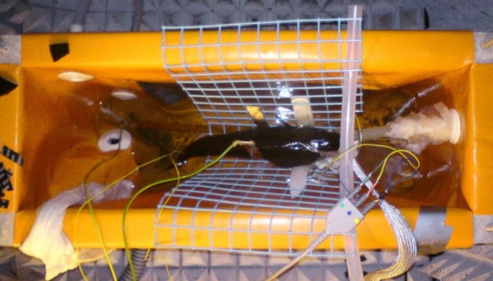
The University of Tromsø, autumn 2012. A cod lies immobilized in a purpose-built special cradle. It has been put to sleep and tubes run oxygenated water through its mouth and gills. Electrodes are attached to its head and tail. The wire on its tail sends electric shocks into the fish. This triggers activity from nerve cells evolved to detect harmful stimuli.
This is called nociception, a reaction common in the animal kingdom, including humans, but distinctive from pain as we know it. Nociception involves the rapid detection of harmful, or noxious, stimulations from all sorts of perils.
Pain, however, entails not only this sensory component but is also a psychological state that includes an unpleasant emotional experience.
Are fish capable of feeling pain, or do their squirms on a fishhook come under the category of nociception? The experiment was devised to see how nociception is recorded and handled by a fish brain.

“This experiment points out a new direction for studying fish and other animals,” says Øyvind Aas-Hansen at the food research institute NOFIMA in Tromsø. He led the research project.
It showed that the cod brain reacts with a surprisingly long time lag, or latency, during and after nociception. In mammals with advanced brains — and in the human brain — this kind of delayed or later reaction to incoming nerve signals is associated with more advanced types of responses.
“Although the latency is longer than previously detected in fish, this study does not determine whether the fish brain is capable of experiencing a feeling such as discomfort with pain, as mammals and humans do,” stresses Aas-Hansen. But he thinks the findings were significant enough to merit further study.
Electrodes under the skin
The new method involves monitoring nerve signals to the entire fish brain. It is far less invasive than previous testing methods, which involved opening the cranium and placing electrodes directly on the brain. Aas-Hansen and colleagues placed electrodes just under the skin at the head of the fish.
“The method is simple and requires no surgery. It enables us to chart responses in the entire brain at once,” explains Aas-Hansen.
Fifteen adult cod were used in the experiment.
Empty stomachs and anaesthetics
The fish had to swim around for five days without being fed prior to the experiment. This is standard procedure in these kinds of tests to ensure that the recording of neural responses are not affected by digestive processes.
“For a cold-water fish this is not as dramatic as it might sound,” says Aas-Hansen.
The Norwegian Animal Welfare Act and other ethical considerations required that the fish be anesthetized. A special medication was selected that would not block the neural impulses to the cods' brains.

The cod were given a muscle relaxant to keep them from flipping about. Any flinches or twitches could also result in conflicting neural impulses.
Tub full of seawater
The fish had to be kept secure during the tests. They were placed in a plastic cradle in a tank of seawater. They were held in place with a metal strap, padded with a moist cloth.
A silicon tube led into the mouth of the fish. It supplied a constant stream of oxygenated seawater through the gills. The water also contained an anaesthetic so the fish would not awaken during the experiment. The water ran from the gills down into a trough below the fish.
Short electric shocks
The fish had to be kept dry on top. Otherwise the electrodes would short circuit in the saltwater. The electrodes were attached subcutaneously in a line running from between the eyes to the fish a centimetre apart.
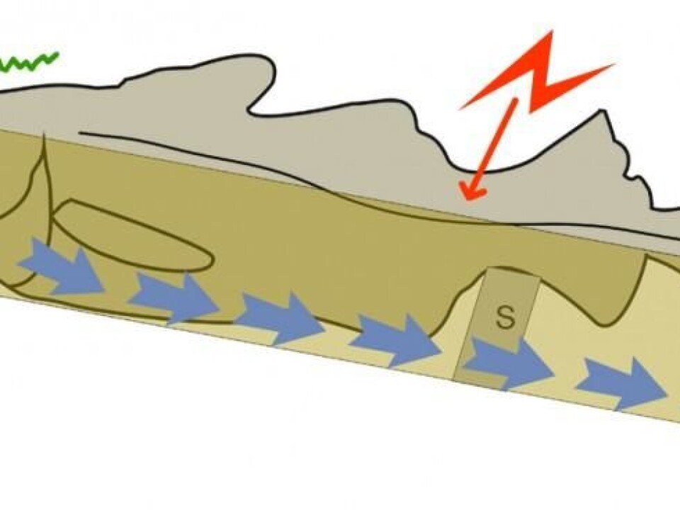
Then two other electrodes were attached to each side of the tail with Velcro. Electrical impulses were sent through these electrodes to instigate pain sensors in the skin of the fish and trigger neural signals to the brain.
The shocks were administered in rapid pulses, a thousand times per second for three seconds. The researchers simultaneously monitored the brains signals from the electrodes on the head of the fish.
Increasing current
The scientists started carefully with a current of just two milliampéres (mA). A human would feel this as a weak vibration in their hand. Then the current was increased fourfold to 20 mA. Most people would be saying “ouch” at this level. But it is not dangerous.
The fish were all anesthetized and consciously felt nothing. That created problems for the Norwegian researchers. If they could have observed a cod trying to flee from the current they could have been more certain that its experience was comparable to what we would suffer.
In any case, they chose to operate with limits that would cause pain in human subjects. The new experimental method resulted in two intriguing observations.
Surprisingly long lag
First of all, the scientists recorded neural signals from the cod up to a quarter of a second after the pulses of electricity were sent. No one has seen that sort of time lag in previous tests.
“Put simply, a delay occurs in the neural signals each time they pass from one neuron to another. The longer time it takes, the more neurons involved along the way,” says Aas-Hansen.
“Our experiment indicates that the most powerful pulses, which most likely initiated nociception, in other words pain pulses in the nerves, are reacted to differently by the cod brain than common stimuli of the senses from the weaker currents,” he said.
Aas-Hansen emphasized, however, that this lag is much shorter than comparable reactions among humans and other mammals.
Many more responses
Secondly, many more responses were detected in the cod brains than in earlier studies with other species of fish.
“This is probably a benefit of this new method, which records the responses in the entire brain at once. We think the high number of responses reflects the fact that the pain reaction is being emitted simultaneously from multiple parts of the brain,” says Aas-Hansen.
This is compelling, because a number of fish researchers have pointed out that the fish brain might react to nociception in different regions than seen in mammals. Birds have also been shown to have alternative parts of the brain involved in this function.
------------
Read the Norwegian version of this article at forskning.no
Translated by: Glenn Ostling